Figure.
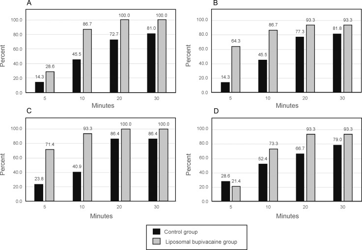
Progression of sensory block. The graphs represent the (A) median nerve, (B) radial nerve, (C) ulnar nerve, and (D) musculocutaneous nerve of the forearm. Data are presented as the percentages of patients with an absence of sensation in the respective nerve at the specified times after the block procedure.

