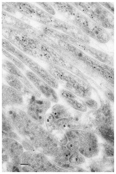
Fig. 5.
Electron microscope immunogold localization of tubulin in the brain of a planarian flatworm. In the top half of the micrograph, there is a bundle of neuron processes with 10 nm immunogold-labeled microtubules; below this is a cluster of synapses. The postsynaptic processes of these synapses lack labeling for tubulin and are spine-like (s). Most of these synapses have two postsynaptic processes. Presynaptic bar or ribbon structures are evident (asterisk). Scale bars is 100 nm. (E7 primary monoclonal antibody to beta tubulin, from Developmental Studies Hybridoma Bank [E7 was deposited to the DSHB by Klymkowsky, Michael (DSHB Hybridoma Product E7)]; specificity of this antibody has been demonstrated previously in a wide range of species (Chu and Klymkowsky 1989, plus numerous references in DSHB); probably Dugesia tigrina, the brown planarian, obtained from Carolina Biological Supplies Company; see Petralia et al. 2010 for general methods, and Petralia et al. 2015 for images from this material showing short invaginating processes in neurons; unpublished data of RSP and Y-XW)

