Fig. 9.
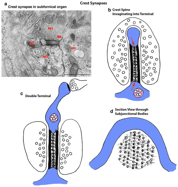
Crest synapses. a Micrograph showing a crest synapse from the subfornical organ (reprint of figure 4A from Akert et al. 1967a; reprinted by permission from Springer Publishing Company). Note the characteristic crest spine with central subjunctional bodies (SB). Other crest synapse structures vary, and in this example, there are two postsynaptic multivesicular bodies (mv; a dark and a light one), and a desmosome-like junction (DS) at the apical bulge of the crest; the presynaptic terminals in this case (pr1 and pr2) contain morphologically different vesicles (red letters have been superimposed over the black letters of the original micrograph, for clarity). Note also that this example is rotated ninety degrees counterclockwise from the drawings in (b–d). b Crest synapse spine invaginating into a single terminal. Red arrows indicate the central plaque of subjunctional bodies between the two PSDs. c Crest synapse spine with a presynaptic terminal on each side of the crest. This example also shows how occasionally a normal spine synapse can project from the top of the crest. d A typical crest synapse that is cut directly through the plane of the central plaque of the subjunctional bodies between the two PSDs, showing the array of subjunctional bodies. The crest synapse often is associated with multivesicular bodies (pink), either in the dendrite near the base of the crest or in the thickened top of the crest spine (Akert et al. 1967a, b; Yamashita et al. 1997) (Color figure online)

