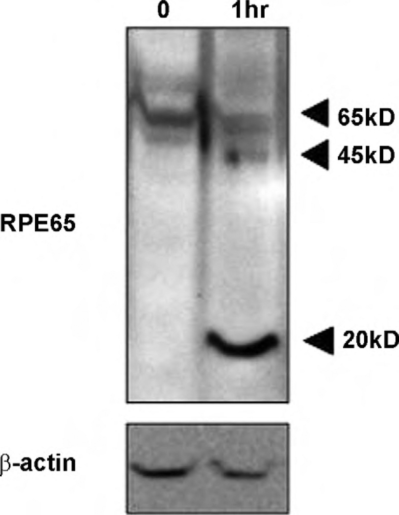
Fig. 1.
RPE45 and RPE20 under oxidative stress in vitro. After 1 h in oxidative stress, RPE65 was cleaved to 45 and 20 kDa fragments (Lane 2), compared to control (Lane 1). RPE D407 cells were treated with 200 µM H2O2 and the control was treated with PBS for 1 h. H2O2 was removed and then cells were maintained in conditioning media for another 6 h before harvest. Proteins were separated by SDS-PAGE (10 µg/Lane), then RPE65 was analyzed by western blot using monoclonal anti-RPE65 protein antibody. RPE cells were maintained in DMEM supplemented with 10% FBS and 2 mM glutamine at 37 °C and 5% CO2.

