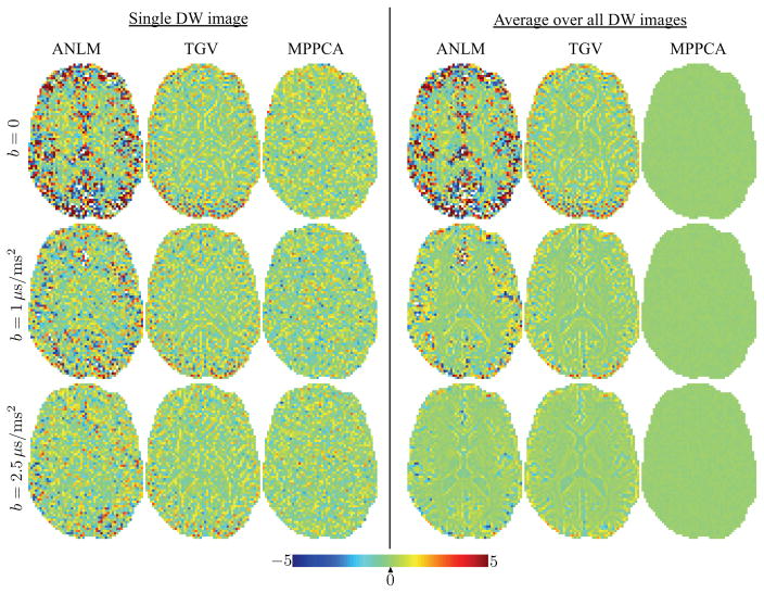
Figure 6.
(left panel) The σ-normalized residual maps between the denoised diffusion-weighted images from Fig. 5 and the original data. (right panel) The presence of anatomical structure in ANLM and TGV anatomical maps indicates interference of the denoising algorithm with the “signal”. The effect becomes more visible after averaging the residual maps of the Mb>0 = 60 images per b value.

