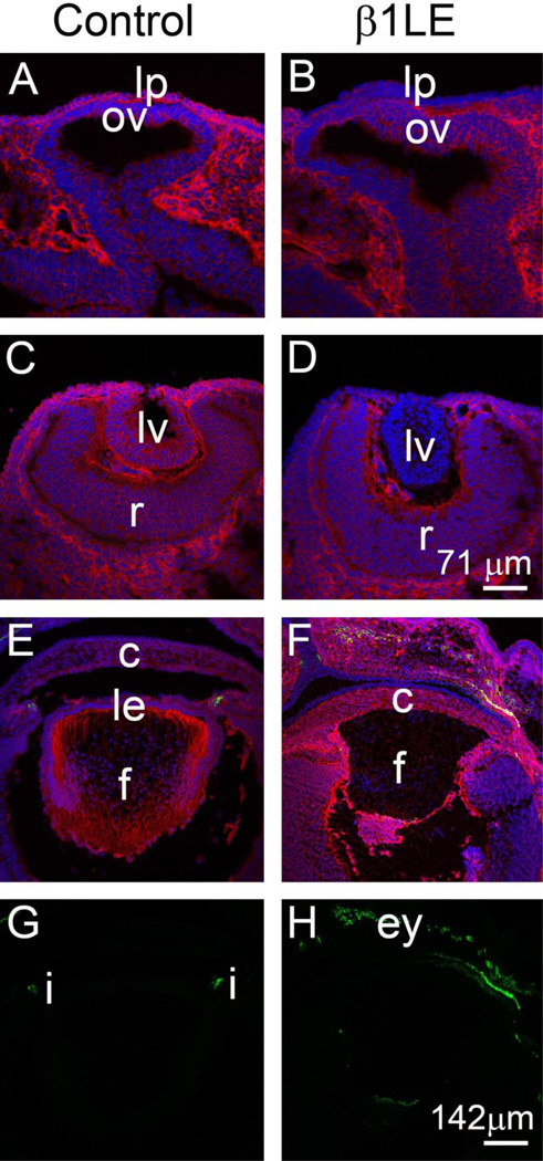
Figure 2. β1LE mice lose β1-integrin protein from the developing lens vesicle by E10.5.
Immunofluorescent confocal microscopy showing β1-integrin protein (red) expression in control (A, C, E) and β1LE (B, D, F) lenses at E9.5 (A, B), E10.5 (C, D) and E16.5 (E, F). At E9.5, β1-integrin protein expression is reduced in the β1LE lens placode (B), compared to control (A). By E10.5, β1-integrin protein is detected in all cells of the lens vesicle of control mice (C) whereas β1-integrin protein levels fall beyond the level of detection in β1LE lenses (D). At E16.5, β1-integrin is still detectable in all cells of control lenses (E) while, consistent with the result at E1 0.5, no β1-integrin was detected in β1LE lenses at this age (F). Co-staining of the E16.5 sections shown in panels E and F for αSMA did not reveal any αSMA signal (green) within the boundary of the lens in either in control (E, see panel G for αSMA channel only) or β1LE lenses (F, see panel H for αSMA channel only). All staining was performed on a minimum of three biological replicates.
Red - β1-integrin, Green - αSMA, Blue - DNA. Abbreviations: lp - lens placode, lv - lens vesicle, le - lens epithelium, c - cornea, f - lens fiber cells, ov - optic vesicle, r - retina, i - iris, ey -eyelids. Scale bars- Panels A, B, C, D-71µm; Panels E, F, G, H- 142 µm

