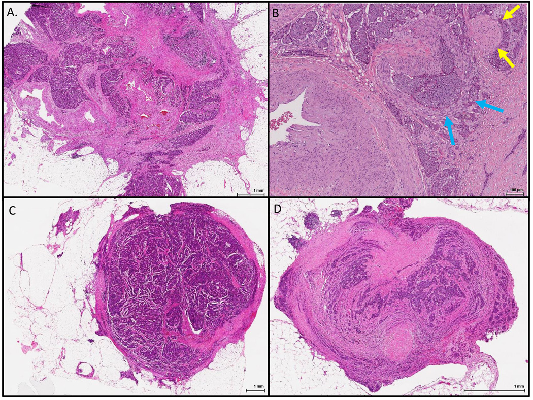
Figure 1.
Representative mesenteric tumor deposits. A. A mesenteric tumor deposit with an irregular contour and associated fibrosis encasing large vessels; B. A higher power view of another mesenteric tumor deposit showing a possible vein involved by tumor (blue arrows) and an entrapped nerve; C. A lymph node completely replaced by neuroendocrine tumor with extra-nodal extension; D. A peritoneal tumor implant with partial mesothelial lining and containing no large vessels.

