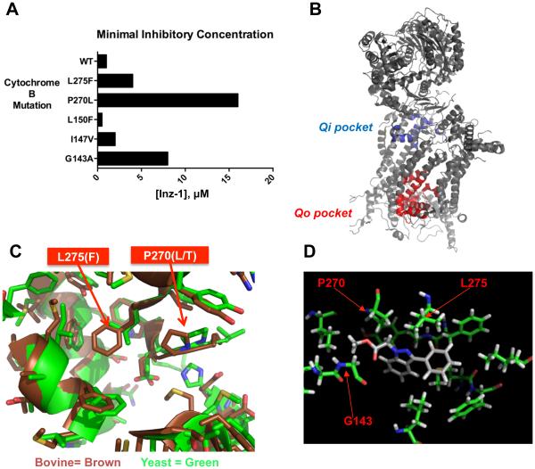
Figure 3. Structural analysis of Inz-1 binding to cytochrome bc1.
A) Minimal Inhibitory Concentration (MIC) of Inz-1 for indicated cytochrome B mutants under respiratory growth conditions in S. cerevisiae. MIC’s were concordant from two independent experiments. B) Structure of yeast cytochrome B complex (PDB:1EZV) indicating location of Qi and Qo pockets. C) Structural alignment of yeast (PDB:1P84) and bovine (PDB:1SQV) cytochrome B showing Qo site and indicating location of two mutations that confer resistance to Inz-1. D) Computational docking of Inz-1 into Qo pocket of yeast cytochrome bc1 (PDB:1EZV) with location of resistance-conferring mutations overlaid.

