
Figure 1.
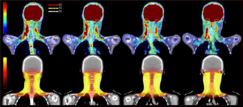
PET CT scan of the neck. Top row – coronal sections showing increased uptake in areas of myositis with superimposed 50 Gy, 55 Gy and 60 Gy isodose lines. Bottom row – corresponding coronal images from radiation treatment planning CT with dose color wash.