Figure 6.
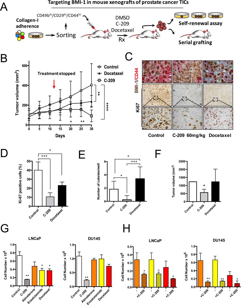
In vivo pharmacological targeting of BMI-1 in mouse PCa xenografts. A, Strategy for examining the antitumor activity of C-209 in serial mouse xenografts and clonogenic repopulation assays of treated cells. B, Growth rate of mouse xenografts generated after subcutaneous (SC) injection of CD49bhiCD29hiCD44hi Luc2EGFP cells. Mice were randomized and administered daily with 60 mg/kg/day of C-209 for twelve days and docetaxel 6mg/kg once a week for two consecutive weeks. Results are mean ± S.D. of six independent experiments. Comparison of tumor volumes between the three groups was determined by two-way ANOVA with Bonferroni post-hoc test. Graph indicates significance of Docetaxel vs. Control at day 30 (**p<0.01) and C-209 vs. Control at day 30 (****p<0.0001). There was a trend towards significance (p=0.08) when comparing tumor volumes in xenograft treated with C-209 vs. Docetaxel at day 30 using Mann-Whitney U test. At the earlier days 20 and 25, Docetaxel was not significantly different than Control, while C-209 was; C-209 vs. Control at day 20 (*p<0.05); C-209 vs. Control at day 25 (**p<0.01). Red arrow indicates treatment discontinuation. In each experiment, n=8/group. C, Intratumor IHC revealed reduced nuclear BMI-1 (brown) and surface CD44 (red) staining upon treatment with C-209. Staining was performed on tumor xenografts taken at Day 30. D, Quantitation of Ki67 positive cells in sections from treated xenografts. (***p<0.001, *p<0.01). E, Colony-forming ability assay performed on freshly dissociated and EGFP sorted xenograft-cells. Average number of colonies/plate for each treatment mean ± S.D. of two independent experiments with 12 wells/condition is reported. *p<0.05, ***p<0.001. F, Tumor initiation potential in serial grafting in secondary mouse xenografts of cells dissociated from treated primary mouse xenografts (n=8 mice /group, *p<0.01). G, Cell proliferation of LNCaP and DU145 cells treated with C-209 (2μM), abiraterone (5μM), enzalutamide (5μM) and docetaxel (2.5nM) for 72 hrs. H, Cell survival of therapy-resistant cells. LNCaP and DU145 cells pre-treated with abiraterone (5μM), enzalutamide (5μM) and docetaxel (2.5nM) for 72 hrs were washed and remaining cells that survived treatments (therapy resistant cells) were treated with C-209 (2μM) for another 3 days. Results are indicated as ± S.D. of two independent experiments with *p<0.05, **p<0.01.

