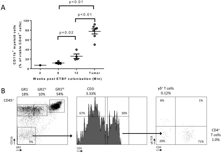
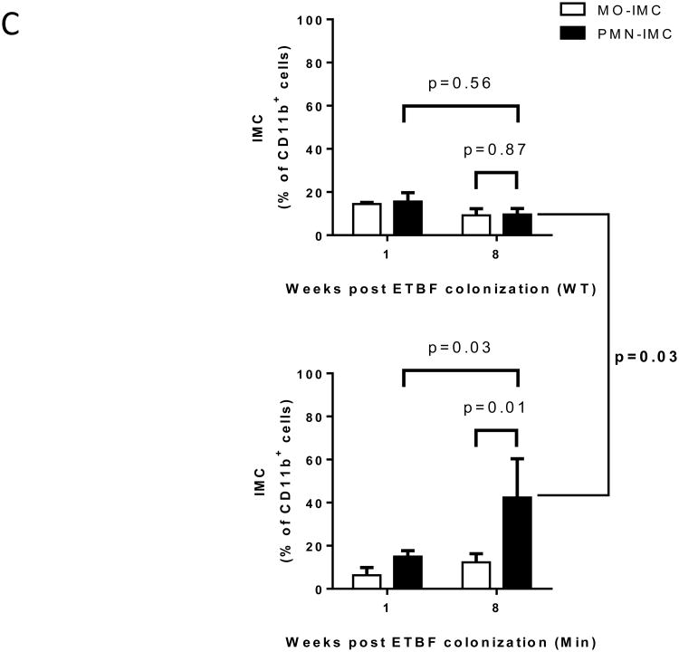
Figure 1. Myeloid cells are the predominant leukocytic population infiltrating ETBF-induced colon tumors in Min mice.
A, Myeloid cells from enzymatically digested grossly normal distal colon tissue (weeks 2, 8 and 12 after ETBF colonization) and distal colon tumors (12 weeks post ETBF) were analyzed by CD45/CD11b staining and flow cytometry. Results are expressed as percent (%) of CD11b+ cells among viable CD45+ leukocytes. Aggregate data of n=7 independent experiments with pooled colon or tumors samples from 3-4 mice/experiment.
B, Flow cytometry of ETBF-triggered colon tumors in Min mice. Percent CD45+ cells are indicated. Representative plots of n=3 or more experiments with 3-4 mice/experiment.
C, Proportion of Ly6ChiLy6G- monocytic immature myeloid cells (MO-IMC) (white bars) and Ly6CloLy6G+ granulocytic (PMN)-IMC (black bars) as percent of CD11b+ isolated from distal colon of WT (top) or Min (bottom) mice at the time points indicated. Of note, IMC in distal colon tissue of sham Min mice were below the limit of detection. Aggregate data of n=4 independent experiments with pooled colon samples from 3 mice/group.


