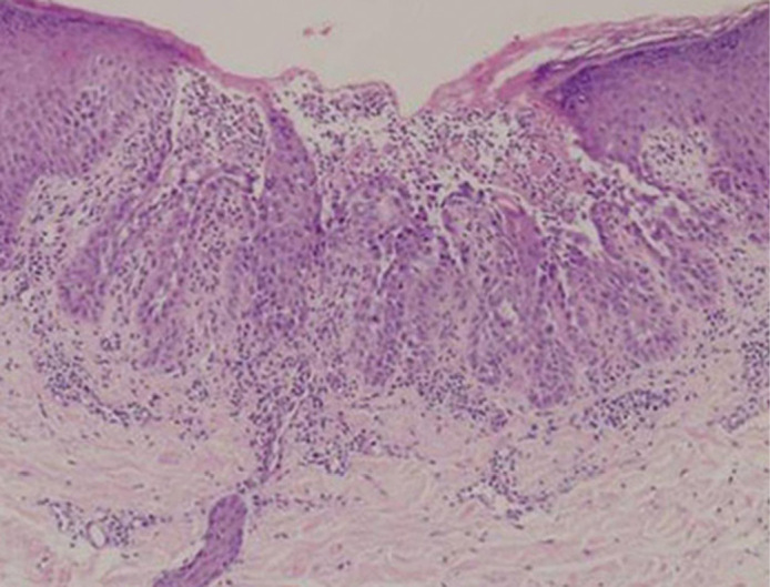
Fig. 2.

Histopathological features of the skin lesions of DD. A skin biopsy specimen from Case 2 shows typical histopathological features of DD, marked hyperkeratosis, acantholysis, and clefts and dyskeratosis in the suprabasal layers of epidermis. Haematoxylin-eosin stain, original magnification X400.
