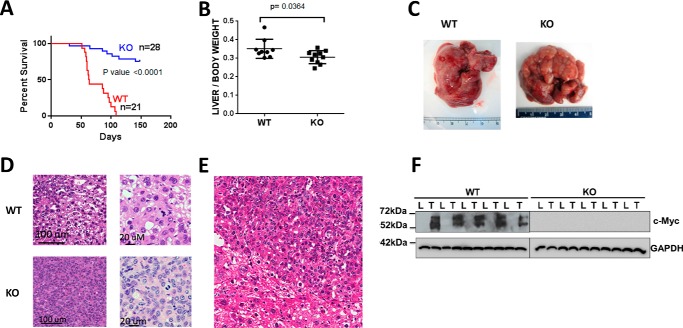
FIGURE 1.
Properties of WT and KO tumors. A, survival curves of WT or KO mice inoculated with mutant β-catenin + YAP SB vectors by HDTVI. The study was terminated at week 22. B, liver weights from the survival curves shown in A. The results include all surviving KO mice. C, gross appearance of typical tumors arising in WT and KO livers. Note that although the depicted WT and KO tumor are of comparable size and appearance, they were obtained at different times due to the slower growth of the latter. D, histologic appearance of WT and KO HBs. Sections were stained with H&E. WT tumors showed small indistinct nodules with variation in nuclear size and frequent mitoses resembling pleomorphic fetal or HCC-like morphology. KO tumors were more distinct with a predominance of small uniform cells with abundant, eosinophilic cytoplasm and uniform small nuclei with inconspicuous to absent nucleoli and many mitoses. E, lower power view of H&E-stained section of normal liver-tumor border emphasizing the size differences between tumor cells (center and upper right) and normal hepatocytes (lower left). The crowded fetal morphology is more apparent. There was also significant nuclear unrest and variation in size in the liver surrounding the nodules. F, immunoblots for Myc protein in livers (L) and HBs (T) from WT and KO mice. GAPDH was included as a loading control. Error bars indicate ± S.E.

