FIGURE 6.
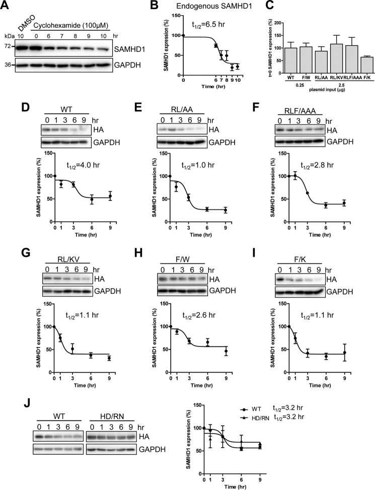
SAMHD1 mutants have decreased half-life compared with WT SAMHD1. Shown are THP-1 or transfected HEK293T cells treated with cyclohexamide (100 μm) and lysates harvested at the indicated times. Immunoblot analysis was used to determine levels of endogenous SAMHD1 protein. A, analysis of endogenous SAMHD1 expression in cyclohexamide treated THP-1 cells at 0, 6, 7, 8, 9, and 10 h post-treatment. The lysates were immunoblotted for SAMHD1 and GAPDH. B, non-linear curve of densitometry from four independent experiments in THP-1 cells. Endogenous SAMHD1 protein half-life (t½) = 6.5 h is indicated. C, comparison of expression levels of WT and mutant SAMHD1 at t = 0 from four experiments used to calculate half-life in D–I. SAMHD1 levels at t = 0 were normalized to GAPDH, and each mutant was calculated as a percentage of WT SAMHD1, which was set as 100%. D–I, HEK293T cells transfected with constructs expressing HA-tagged WT or mutant SAMHD1 to normalize protein expression to equal levels. At 48 h post-transfection, cyclohexamide treatment was started, and lysates were collected at the indicated time points post-transfection. Lysates were immunoblotted for HA and GAPDH. D–I, non-linear curves, t½ calculated from immunoblotting densitometry, and one representative immunoblot. Densitometry data and t½ was calculated from a minimum of four independent experiments. J, comparison of WT and the dNTPase defective HD/RN mutant (H206R and D207N) showing immunoblotting analysis (left panel) and non-linear curves with calculated t½ (right panel). The data represent two independent experiments.

