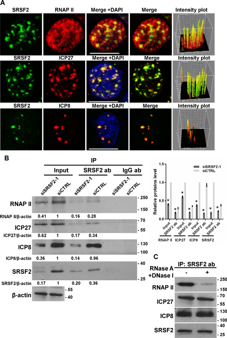
FIGURE 4.
SRSF2 associated with RNAP II, ICP27, and ICP8. A, after 4 h of infection with HSV-1, HeLa cells were fixed, stained with antibodies against SRSF2 (green), RNA polymerase II (red), ICP27 (red), and ICP8 (red), and subjected to confocal microscopy analysis. The intensity plots for the red and green channels were analyzed with ImageJ software. DAPI (blue) was used to stain the nuclei. Scale bars, 10 μm. B, after 36 h of transfection with the SRSF2 siRNA or negative control siRNA, HeLa cells were infected with HSV-1 at a m.o.i. of 1 for 4 h. Cell lysates were harvested and subjected to an immunoprecipitation (IP) assay with the anti-SRSF2 antibody or the anti-IgG antibody (Ab). The retrieval of RNA polymerase II, ICP27, SRSF2, and ICP8 by endogenous SRSF2 and IgG was measured by Western blotting. Protein ratios for the indicated proteins/β-actin were analyzed with ImageJ software, and statistical analysis was conducted with data from three independent experiments (right panel). The data are presented as the mean ± S.D. (*, p < 0.01, Student's t test). C, HeLa cells were infected with HSV-1 at a m.o.i. of 1 for 4 h. Cell lysates were harvested with and without DNase and RNase treatment and then subjected to an immunoprecipitation assay with the anti-SRSF2 antibody. The retrieval of RNA polymerase II, ICP27, SRSF2, and ICP8 by endogenous SRSF2 was measured by Western blotting.

