Figure 5.
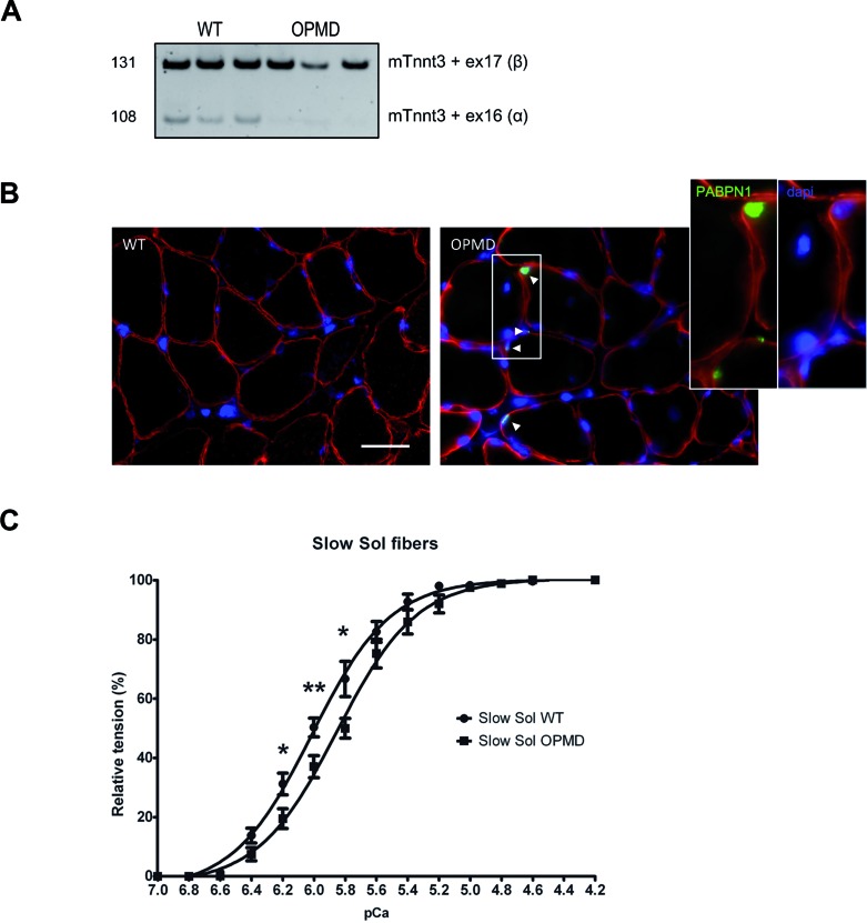
Splicing defect in OPMD mouse induces decreased calcium sensitivity. (A) Tnnt3 splicing defect is present in OPMD mouse model. RT-PCR analysis of endogenous Tnnt3 exons 16 and 17 isoforms in soleus (Sol) muscles of wild-type (WT) and A17.1 (OPMD) mice. (B) PABPN1 staining following KCl treatment revealed intranuclear aggregates in the soleus muscle of A17.1 mouse (red: dystrophin, green: PABPN1, blue: dapi), whereas no aggregate are observed in control mice. Aggregates are indicated with an arrow (see also inset boxes). Scale bar = 30 μm. (C) Deregulation of Tnnt3 exon 16 and 17 induces decreased calcium sensitivity in mice slow fibers, as measured by pCa/Tension relationship. * indicates P< 0.05, ** indicates P< 0.01.

