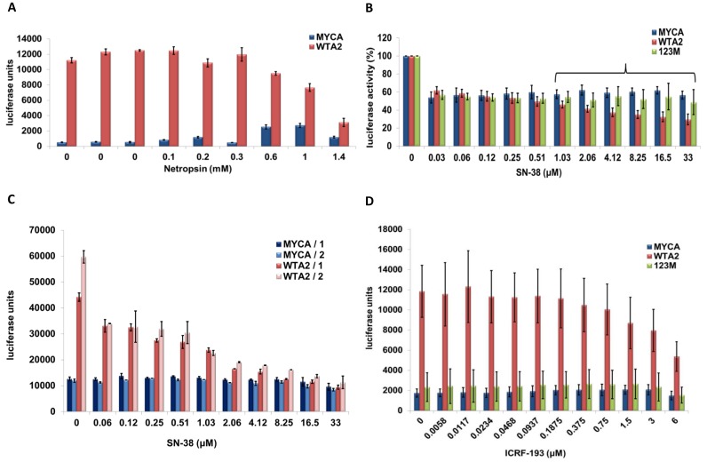
Figure 3.
Netropsin, SN-38 and ICRF-193 are HMGA2 antagonists. (A) Representative luciferase reporter assays of one of three sets of co-transfections with mock or wild-type HMGA2. Cells were exposed to increasing concentrations of netropsin, as indicated, for 24 h before luciferase assays were performed. We show mean values plus standard deviations of readings from four replicates for each netropsin concentration and for three sets of four replicates without netropsin as controls. (B) Luciferase reporter assays were performed with HeLa cells for six independent sets of co-transfections with mock, wild-type HMGA2 or mutant HMGA2 that carried substitutions in all three AT-hooks (123M), as described in the text. In each case, cells were exposed to increasing concentrations of SN-38 for 24 h, and data from two replicates per co-transfected pair of vectors were collected. Mean values from each set of co-transfections were normalized to SN-38-untreated controls. We show the mean values of the combined six normalized data sets with standard deviations. The bracket indicates when statistically significant (P < 0.05) differences between mock and wild-type HMGA2 as well as between mutant HMGA2 and wild-type HMGA2 readings could be determined; Student's t-test. (C) Luciferase reporter assays were performed with H1299 cells for two independent co-transfections with either mock (MYCA) or wild-type HMGA2 (WTA2) in combination with Renilla expression vector. Cells from each co-transfection were plated 24 h later in two rows of 11 wells on a 96-well plate. The next day, SN-38 was titrated in each row, and luciferase activity was measured after additional 24 hours. The bars show mean values with standard deviations from these two luciferase measurements for each co-transfection. (D) Luciferase reporter assays were performed with HeLa cells with mock (MYCA), wild-type HMGA2 (WTA2), or HMGA2 mutant (123M) in combination with Renilla expression vector. Two independent sets of co-transfections were performed. In each set, cells from two six wells (e.g. mock plus Renilla vectors) were combined 24 h after co-transfection and plated in three rows of 12 wells on a 96-plate. Each triplicate of rows was repeated three times, and ICRF-193 was titrated in 24 h later. The bars show mean values with standard deviations from the combined 18 luciferase measurements obtained for each co-transfection after one day of drug exposure.

