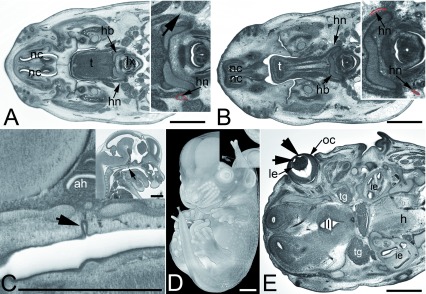
Figure 4. Other frequently observed abnormalities in mutant embryos.
A and B. Abnormal hyopglossal nerve in original HREM sections through the head of Prrc2b -/- ( A) and a Polb -/- ( B) embryo. Note the missing right hypoglossal nerve (arrowhead, inlay) in A and the thinning of both hypoglossal nerves (hn) in B. C– E. Abnormalities that also occur in controls. Persisting craniopharnygeal duct (arrowhead) as appearing in sagittal sections ( C). Split tip of tail featured by volume models ( D) and vesicles (arrowheads) in the lens (le) as appearing in an original HREM section ( E).

