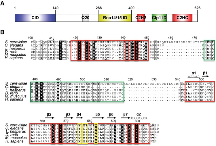
FIGURE 1.
(A) Domain organization of S. cerevisiae Pcf11, drawn with IBS (Liu et al. 2015). (B) Sequence alignment versus secondary structure for the C terminus of Pcf11 from S. cerevisiae to H. sapiens, drawn with ESPript (Robert and Gouet 2014). Conserved residues are indicated in black or in bold, based on similarity levels. Two putative zinc fingers and Clp1 interacting regions are highlighted in red boxes and a green box, respectively. Zinc coordinating Cys and His residues are highlighted in red boxes. Mutated residues Y575, E577, E578, and W582 are highlighted in yellow boxes.

