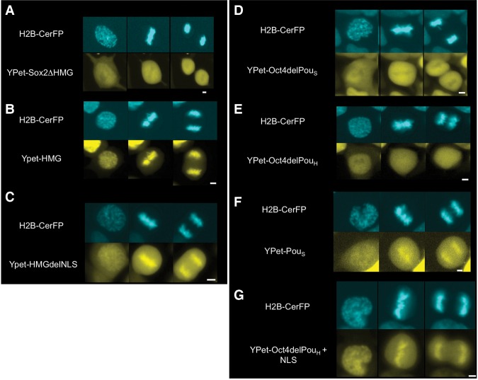
Figure 2.
MCB of SOX2 and OCT4 is mediated by their HMG and POUS domains, respectively. Fluorescence snapshots of living ES cells expressing H2B-CerFP and dox-inducible YPet fusion to truncated versions of SOX2 and OCT4. (A) SOX2 with a deletion of its HMG DNA-binding domain. (B) HMG DNA-binding domain of SOX2. (C) HMG DNA-binding domain of SOX2 without its NLS. (D) OCT4 with a deletion of its POUS DNA-binding domain. (E) OCT4 with a deletion of its POUH DNA-binding domain. (F) POUS DNA-binding domain of OCT4. (G) OCT4 with a substitution of its POUS DNA-binding domain with a NLS. In each panel, prophase is at the left, metaphase is in the middle, and anaphase is at the right. Bars, 5 µm.

