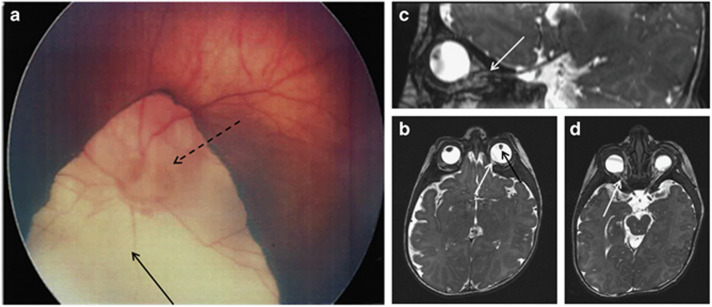
Figure 2.
Fundus and brain MRI results. (a) Fundus view showing the retinal coloboma (full-line arrow) of left eye involving the optic disk (dotted-line arrow) – type IV of the Gopal classification. (b–d) Brain and optic nerve MRI (2 months) showing bilateral microphthalmia (b) associated with irido coloboma (black arrow) and retinal coloboma (white arrow), and the bilaterally thin (hypoplastic) optic nerve hypoplasia predominantly in the intra-orbital portion (c, d) (white arrows).

