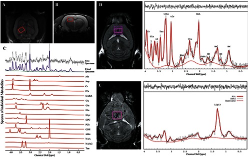
Figure 2.

In vivo T2-weighted MR images A) coronal and B) axial showing regions of interest (ROI) for 1H-MRS experiments. Here, the ROI is centered in the right dorsal hippocampal region of the rat brain. C) Quantification of neuro-metabolite signals from in vivo 1H-MRS in the right dorsal hippocampal region of the Stress-induced Sleep Perturbation (SSP) model. D) 1H-MRS voxel location on a T2-weighted MRI and the corresponding 1H-MRS spectra from a control mouse and E) from a mouse with large a glioblastoma. 1HMRS spectra from glioblastoma are characterized by increased lipid signals. tCr, total creatine; Glx, glutamate + glutamine; Ins, myo-Inositol; Tau, Taurine; tCho, total choline; NAA, N-acetyl-aspartate; Glu, glutamate; MM, macromolecules; Lac, Lactate; GABA, gamma-aminobutyric acid. A, B and C panels were adapted from Lee et al., PLoS One 2016;11:e0153346 with permission; D and E panels were adapted from Park et al., PLoS One 2014;9:e94755 with permission.
