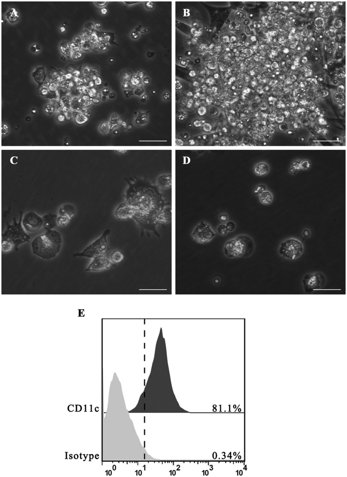
Figure 1. Morphology of chicken BM-DCs cultured with recombinant chicken GM-CSF and IL-4.
(A) Light microscopy showed cell aggregates after 4 days of culture (100×). (B) The size of these aggregates increased after 24 h of stimulation with B.S. or B.S.-HA (100×). (C) Morphology of cells after 7 days of culture after stimulation with B.S. or B.S.-HA (400×). (D) Culture of chicken bone marrow cells without chicken GM-CSF and IL-4 (100×). Scale bars =20 μm. (E) More than 81% of immature chicken BM-DCs were CD11c+ by flow cytometry.

