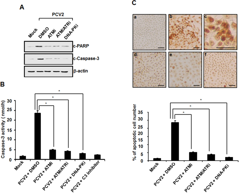
Figure 6. Inactivation of PCV2 stimulation of the DDR pathway suppresses virus-mediated apoptotic responses in host cells.
(A) Monolayers of PK15 cells were infected with PCV2 in the presence or absence of the DDR inhibitors. At 48 h postinfection, the cell lysates were collected for Western blot analysis with specific antibodies to detect the cleavage of PARP and caspase-3. Equal protein loads were verified with β-actin blots. p-, phosphorylated; c, cleaved. (B) Whole-cell lysates collected from the PCV2-infected cells at 48 h after treatment with the DDR inhibitors were assayed for DEVDase activity by using the caspase-3 colorimetric DEVD-AFC. Furthermore, the cells infected with PCV2 alone were treated with DEVD-CHO, a caspase-3 inhibitor. Mock-infected cells were acted as a negative control. Values expressed are means from three independent experiments. (C) Inhibition of DDR activation reduces apoptosis caused by PCV2 infection by TUNEL staining. The PCV2-infected cells at 48 h after treatment with the DDR inhibitors were processed for the TUNEL assay. Upper panel, shown is TUNEL staining for fragmented DNA. The cells were mock infected (a), and infected with PCV2 in the absence (b,c is partial enlargement of b) or the presence of ATM (d), ATM/ATR (e), or DNA-PK inhibitor (f). Brown dots are TUNEL-positive nuclei. Bars, 80 μM. Data are representative of data from an experiment performed in repeated three times with similar results. Lower panel, TUNEL positivity was determined as the average percentages of apoptotic nuclei. Data are means ± SD from three independent experiments. *P < 0.05 for a comparison of the PCV2-infected and PCV2-infected DDR inhibitor-treated cells.

