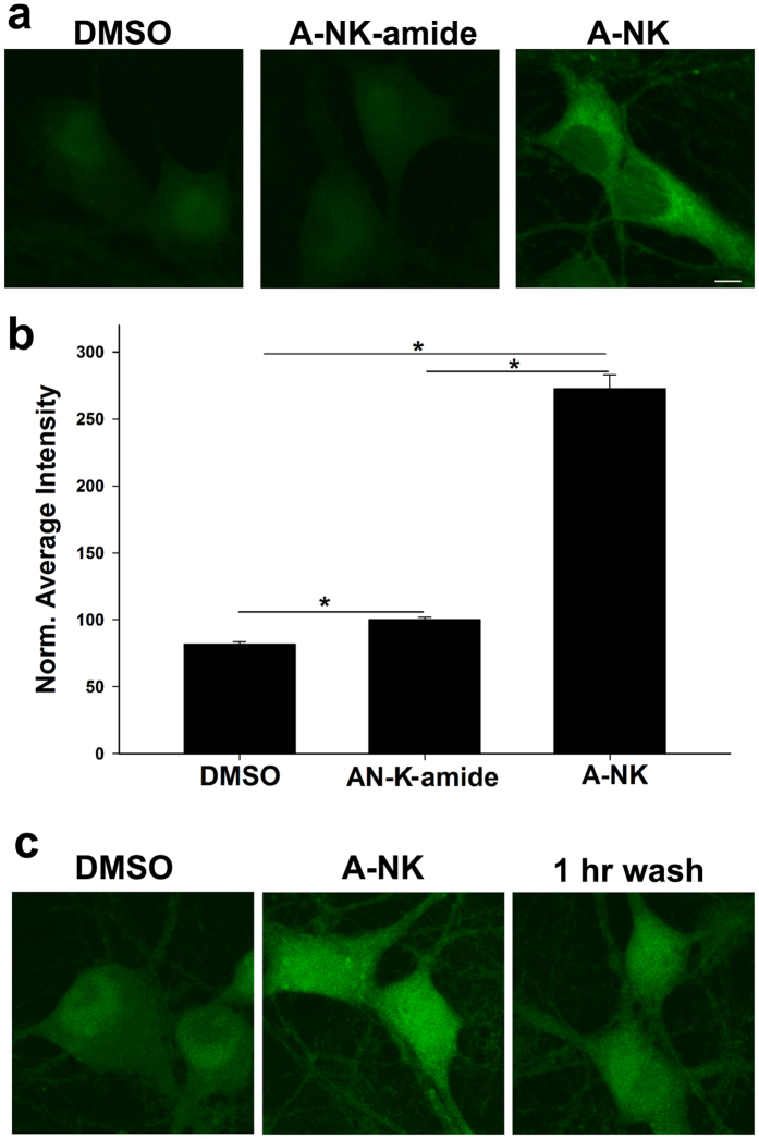
Figure 7. A-NK but not A-NK-amide strongly accumulates inside neurons.
(a) Confocal imaging depicting cells exposed to DMSO, 20 μM A-NK-amide or 20 μM A-NK, permeabilized and stained with azide-Alexa Fluor 488 under copper-catalyzed “click” reaction conditions. Labeling is observed in the A-NK treated dish but is much weaker for A-NK-amide. Scale bar: 5 μm. (b) The amount of fluorescence in each condition was quantified (n = 15 neurons from 3 separate experiments, *p < 0.05, paired t-test). (c) Cells were incubated in 10 μM A-NK for 1 h, followed by fixation. Cells were either processed immediately for click cyto-fluorescence or were washed for 1 h in phosphate-buffered saline before processing. Images are representative of three independent experiments. Scale bars in A and C: 5 μm.

