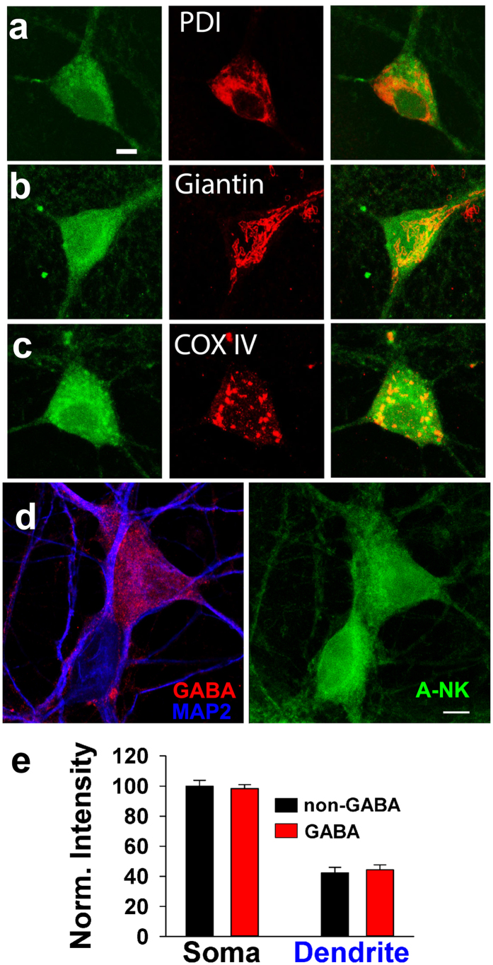
Figure 8. A-NK staining involves multiple intracellular compartments.
(a–c) Left panels depict confocal imaging of staining of cells exposed to 10 μM A-NK, permeablized and stained with azide-Alexa Fluor 488 under copper-catalyzed “click” reaction conditions. Middle panels show immunofluorescence staining of the ER (anti-PDI, a), Golgi (anti-Giantin, b) or mitochondria (anti-COXIV, c). Right panels are merged images indicating that the A-NK staining did not overlay specifically with any of the organelle markers. Scale bar: 5 μm. (d) To test for preferential A-NK accumulation in dendrites and interneurons, we co-labeled with antibodies for GABA (red) and MAP2 (blue). The right panel shows A-NK accumulation. (e) Summary of A-NK fluorescence from confocal projections in somas and dendrites of interneurons (n = 19) and principal neurons (n = 17).

