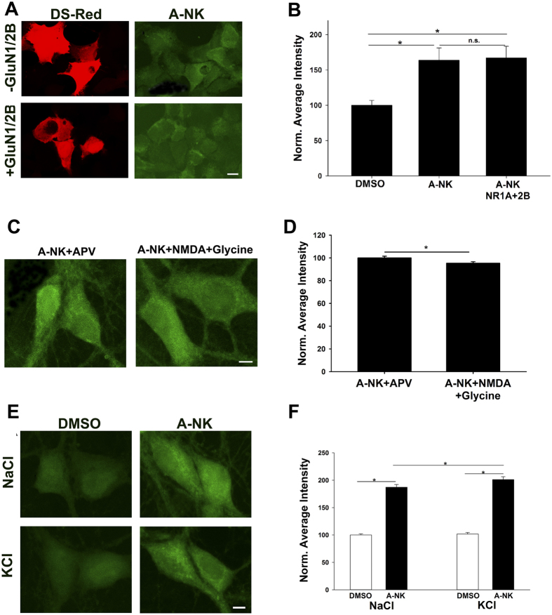
Figure 9. A-NK accumulation does not require NMDAR presence.
(a) HEK cells were transfected with either DsRed alone or with NMDAR subunits GluN1a and GluN2B as indicated. Cells were incubated with 10 μM A-NK for 15 min, then fixed and processed for click cyto-fluorescence. Scale bar: 5 μm (b). Quantification of fluorescence intensity from 5 independent experiments relative to sibling negative control dishes (DMSO treated; *p < 0.01). (c,d) Channel activation does not increase A-NK labeling in neurons. (c) Images of neurons co-incubated in 50 μM D-APV to prevent channel activation during A-NK exposure or in co-agonists 10 μM NMDA and 10 μM glycine to activate channels (1 h incubations). Scale bar: 5 μm. (d) Summary from 40 neurons in 4 independent experiments showing that channel activation slightly but significantly decreased A-NK accumulation (*p < 0.05), in contrast to expectations that permeation of the NMDAR may facilitate A-NK entry. (e,f) Depolarization does not reduce A-NK entry. (e) Neurons were incubated in 10 μM A-NK in normal saline or in saline containing 120 mM KCl to reduce the resting membrane potential to near 0 mV. Scale bar: 5 μm. (f) Summary of A-NK accumulation from 30 neurons in 3 experiments. Depolarization slightly increased A-NK accumulation (*p < 0.01), opposite of an electro-attraction mechanism of entry and retention.

