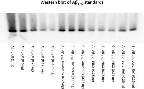
Fig. 2.

Western blot of amyloid-β 1–42 peptide (Aβ1–42) peptide standards after 3D6 antibody detection following quantitative acid gel electrophoresis. Lanes 1–5 (left to right),1–42 reference standard from Lilly with amounts of 1, 0.5, 0.25, 0.125, and 0.0625 ng, respectively, loaded into each well. Lanes 6–8 show three replicates of the diluted Quanterix Aβ1–42 standard. Lanes 9–11 show three replicates of the diluted INNOTEST® Aβ1–42 standard. Lanes 12 and 13 show two replicates of another Lilly substock of Aβ1–42 reference standard. From lane 6 to lane 13, 0.25 ng of each Aβ1–42 peptide was loaded per well, based upon their stated concentrations
