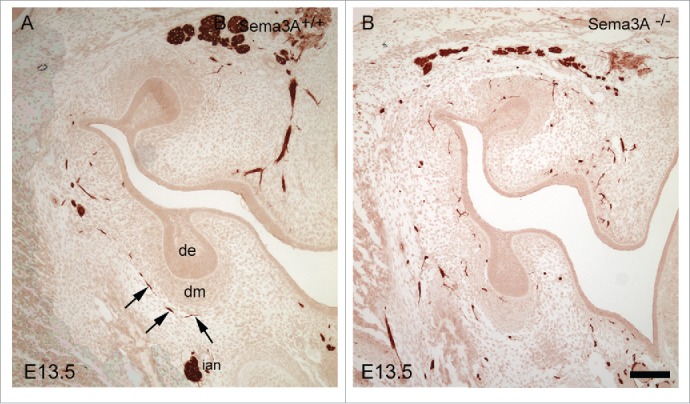
Figure 2.

Immunohistochemical localization of nerve fibers in Sema3A+/+ (A) and Sema3A−/− (B) bud stage molar tooth germs at E13.5. Nerves in the Sema3A−/− molars and mandible mesenchyme exhibit apparent defasciculation as well as abnormal patterning and tooth target innervation (e.g. ectopic expression in the condensed dental mesenchyme and next to the dental epithelium). In contrast, in the Sema3A+/+ tooth, germ dental nerves show organized, appropriate target innervation (arrows). Abbreviations: cm, condensed dental mesenchyme; de, dental epithelium. Scale bar: 100 µm.
