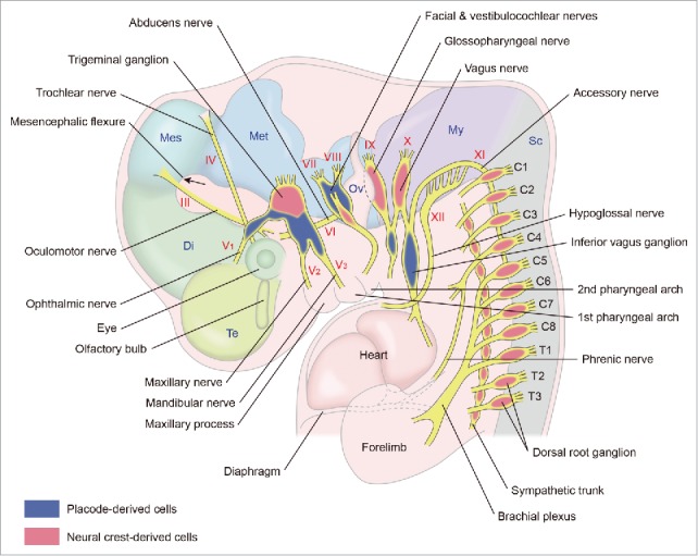
Figure 1.

A schematic lateral view of the peripheral nervous system in a higher vertebrate embryo at the early stages. The 3rd–12th cranial nerves with their ganglia, spinal nerves with dorsal root ganglia and sympathetic trunk with its ganglia are shown. C1–C8 and T1–T3 indicate 1st–8th cervical and 1st–3rd thoracic spinal nerves, respectively. Di: diencephalon, Mes: mesencephalon, Met: metencephalon, My: myelencephalon, Ov: otic vesicle, Sc: spinal cord, Te: telencephalon.
