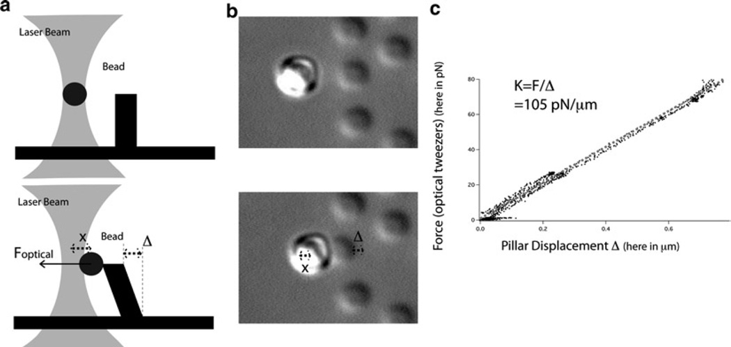
Fig. 4.
Calibration of the pillars using optical tweezers. (a) Schematics of the calibration of the pillars using optical tweezers: at the beginning of the calibration when the bead is not in contact with the pillar and then in the middle of the calibration when force is exerted on the pillar. (b) Optical microscopy images of the calibration of the pillars using optical tweezers: at the beginning of the calibration when the bead is not in contact with the pillar and then in the middle of the calibration when force is exerted on the pillar. (c) An example of the calibration of the pillars with optical tweezers: the slope of the graph of the force of the optical tweezers vs. the displacement of the pillar gives the stiffness of the pillar.

