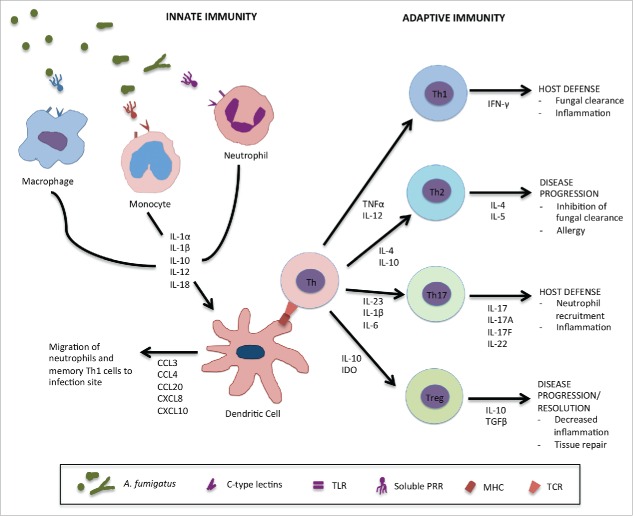
Figure 1.
Host immune response against A. fumigatus. Conidia which enter the lungs and escape airway epithelial cells are recognized by innate immune effector cells via cell surface pattern recognition receptors (PRRs) such as dectin-1 or toll-like receptors (TLRs) and soluble factors such as pentraxin-3 (PTX-3), surfactant proteins A and D (SP-A,D) or mannose-binding lectin (MBL). Alveolar macrophages recognize and clear conidia leading to the activation of inflammatory pathways and the secretion of various proinflammatory cytokines by epithelial cells, other macrophages and dendritic cells. The cytokine environment contributes to the recruitment of other innate immune effectors such as inflammatory monocytes and neutrophils which mediate fungal killing of conidia that have escaped initial recognition and germinated into hyphae and further stimulate proinflammatory responses. Dendritic cells (DCs) are the link between the innate and adaptive immune responses to A. fumigatus. During infection and after interaction with other innate effectors, they secrete various chemokines such as CCL3, CCL4, CCL20, CXCL8 and CXCL10 to allow migration of more inflammatory cells to the site of infection. They also interact with immature helper T cells (Th) through the major histocompatibility complex (MHC) and T cell receptor (TCR), resulting in the secretion various cytokines driving protective Th1 and Th17 responses against A. fumigatus. They can also inhibit Th2 responses which favor disease progression and allergy. Regulatory T cells are producers of IL-10 which is linked to disease progression in IA.

