Figure 1.

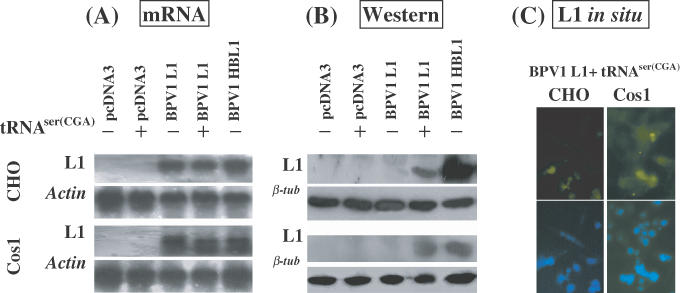
Translation of BPV1 L1 mRNA is enhanced by provision of tRNASer(CGA). CHO or Cos1 cells were transiently transfected with (i) pCDNA3, (ii) pCDNA3 + pSVtRNASer(CGA), (iii) pCDNA3BPV1 L1, (iv) pCDNA3BPV1 L1 + pSVtRNASer(CGA) and (v) pCDNA3BPV1 HBL1 (codon modified). (A). Northern blot hybridization of L1 mRNA transcription. RNA samples were prepared at 42 h post-transfection. Of each sample of DNase-I-digested total RNA, 10 μg was electrophoresed on a 1.2% denatured agarose gel and blotted onto a nylon membrane. The northern blots were hybridized with 32P-labeled wt and HB L1 probe mixture. As controls, the northern blots were hybridized with 32P-labeled actin gene probe. (B) Immunoblotting analysis of the L1 protein. Monoclonal antibody against BPV L1 protein was used to probe the blots. Upper panels show the results of L1 immunoblotting assay; lower panels show the results of β-tubulin immunoblotting assay indicating equal loading of the protein samples. (C) L1 expression in CHO and Cos1 cells co-transfected with pCDNA3BPV1 L1 and pSVtRNASer(CGA) was demonstrated by indirect immunofluorescence microscopy. Upper panels show the L1 labeling; lower panels show the nuclear staining by DAPI.
