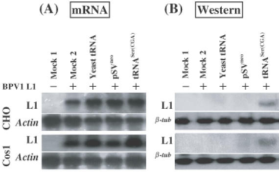
Figure 2.

Enhancement of translation of BPV1 wt L1 mRNA by tRNASer(CGA) is specific. CHO or Cos1 cells were transiently transfected with (i) pCDNA3 (mock 1), (ii) pCDNA3BPV1 L1 (mock 2), (iii) pCDNA3BPV1 L1 plus exogenous supplement of yeast tRNAs in culture medium (commercial bake yeast tRNAs from Sigma, Australia, were added into CHO or Cos1 cell cultures at 1 μg/ml following transfection of BPV1 wt L1 expression construct), (iv) pCDNA3BPV1 L1 plus pSVneo basal vector, and (v) pCDNA3BPV1 L1 plus pSVtRNASer(CGA). (A). Northern blot hybridization of L1 mRNA transcription. RNA samples were prepared at 42 h post-transfection. Of each sample of DNase-I-digested total RNA, 10 μg was electrophoresed on a 1.2% denatured agarose gel and blotted onto a nylon membrane. Northern blots were hybridized with 32P-labeled BPV1 wt L1 probe (upper panel). As controls, the northern blots were also hybridized with 32P-labeled actin gene probe (lower panel). (B) Western blotting analysis of L1 protein. Monoclonal antibody against BPV L1 protein was used to probe on the blot. Upper panels show the results of L1 immunoblotting assay; lower panels show the results of β-tubulin immunoblotting assay indicating equal loading of the protein samples.
