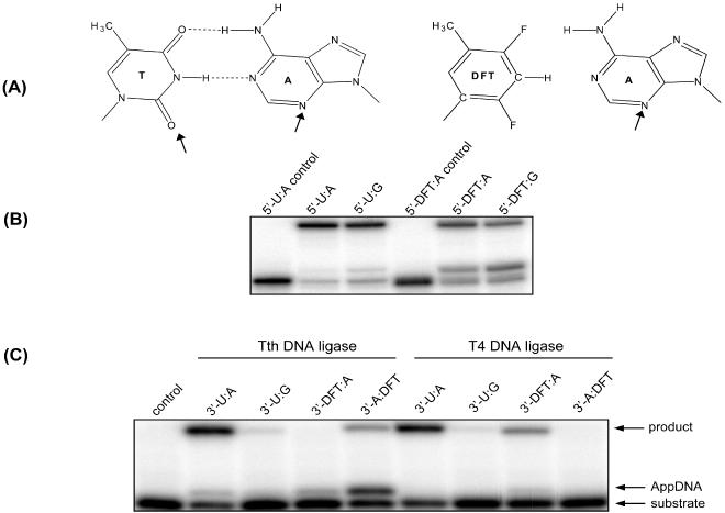
Figure 6.

DNA ligase assays with substrates containing DFT. (A) The structure of DFT is shown above the gel, and hydrogen bonding acceptors in the minor groove of an A:T pair are indicated by arrows. (B) Tth DNA ligase assays with DFT on the 5′-phosphate end of a ligase junction. The experiments were conducted at 26.5°C for 30 min with 0.5 pmol of duplexes and 10 U of Tth DNA ligase in a total volume of 10 μl. Control lanes contain no protein. (C) Tth and T4 DNA ligase assays with DFT on the 3′-hydroxyl end of a ligase junction. Both ligase experiments were conducted with equimolar DNA duplex and enzyme (100 fmol each) in a total volume of 20 μl. Tth ligase reactions were incubated at 26.5°C for 5 min, and T4 ligase at 16°C for 10 min in the corresponding reaction buffer as described in Materials and Methods. 3′ (5′)-X:Y indicates that X is on the 3′-hydroxyl (5′-phosphate) end of the ligase junction, and Y is on the template.
