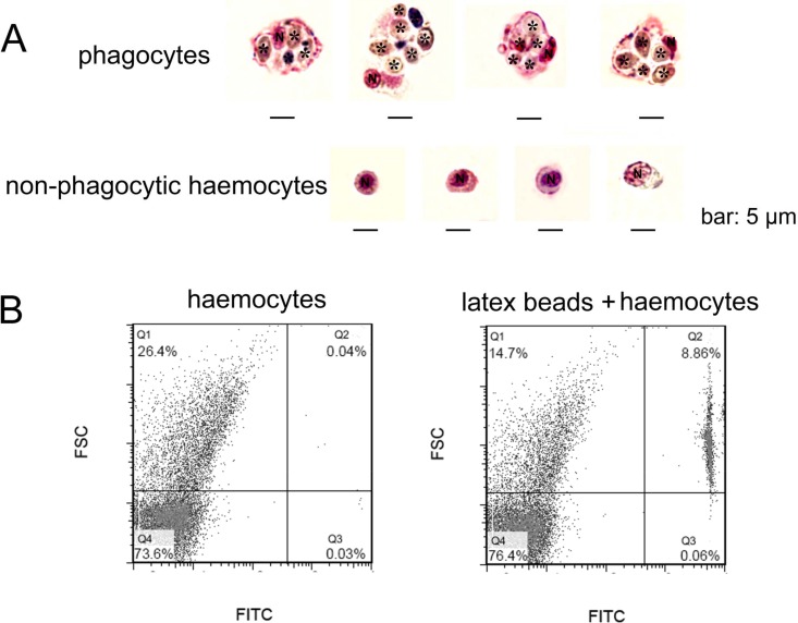
Figure 2. Morphological identification of phagocytes from C. gigas.
(A) Haemocytes were incubated with fungal cells Pichia pastoris to allow phagocytosis, followed by MGG staining and microscopic analysis. Fungal cells are indicated with asterisks, N stands for cell nucleus. Bar: 5 µm. (B) Haemocytes pre-incubated with FITC-labeled latex beads (2 µm diameter) and analyzed by flow cytometry.

