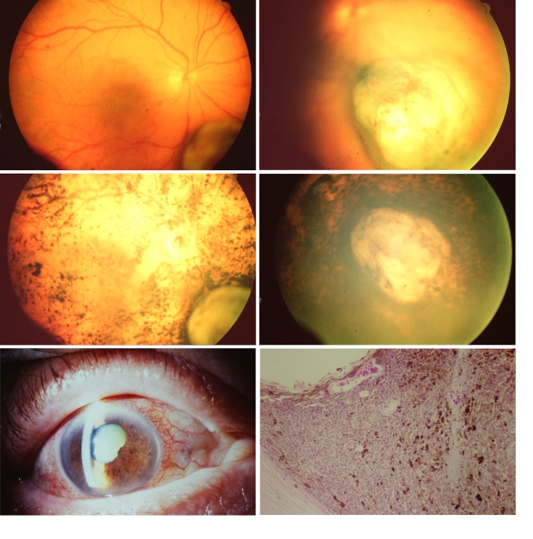
FIGURE 1.
Clinical photographs of the eye and the choroidal melanoma that had been treated with an iodine-125 plaque in August 1988 and after enucleation gave rise to cell line Mel202. Top row, fundus and choroidal melanoma at the time of diagnosis in July 1988. Middle row, fundus and choroidal melanoma in January 1989 (left) and in March 1989 (right). Bottom row, eye just prior to enucleation in 1990 (left) and histologic section of the scleral part of the tumor (right), illustrating the high density of blood vessels and pigment macrophages (hematoxylin-eosin, ×10).

