Figure 2.

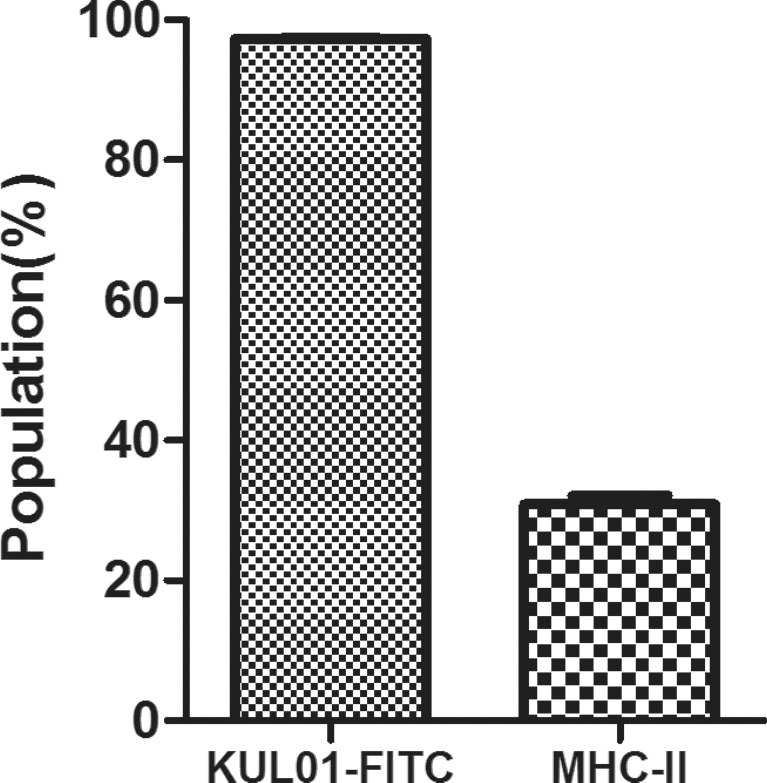
Expression of surface molecules on chicken MDM cells. Cultured MDM were stained with monoclonal antibody KUL01-FITC and MHC-II and analyzed using flow cytometry.

Expression of surface molecules on chicken MDM cells. Cultured MDM were stained with monoclonal antibody KUL01-FITC and MHC-II and analyzed using flow cytometry.