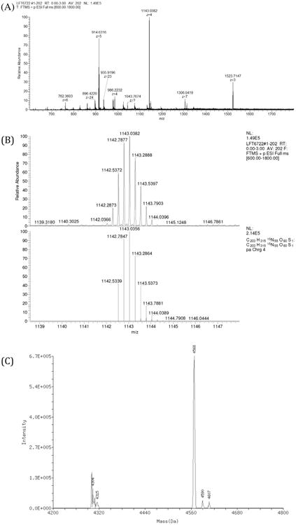
Fig. 4.

An ESI mass spectrum of the HPLC-purified 15N Aβ42 peptide in positive ion mode. (A) Full spectrum depicting various charge states of 15N isotope-labeled Aβ42 peptide. (B) Isotopic distribution of +4 charged species (top panel) and theoretical simulation of +4 charged species generated from 15N Aβ42 peptide in Xcaliber (bottom panel). (C) A deconvolution of the peaks from full spectrum to generate monoisotopic mass of the 15N Aβ42 peptide.
