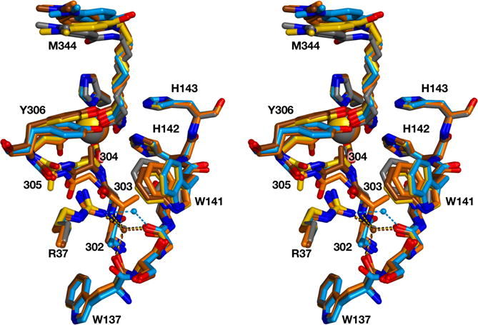
Figure 4.

Stereoview of the glycine-rich loop of each structure superimposed on that of wild-type HDAC8 complexed with M344 (grey; PDB ID: 1T67). Each structure is color-coded as in Figure 3: G302A HDAC8, blue; G303A HDAC8, orange; G304A HDAC8, brown; G305A HDAC8, yellow. For reference, R37 and the W137–W141 segment are also illustrated, and the catalytic Zn2+ ion is a large sphere. The bridging water molecule near R37 is shown as a small sphere and hydrogen bonds are shown as dotted lines.
