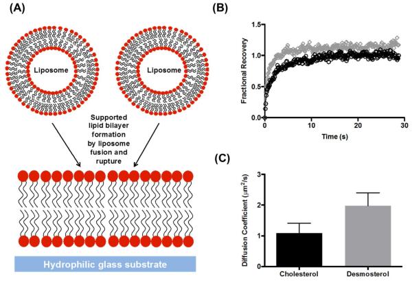
Figure 4. Effect of desmosterol on SLB mobility.
(A) SLBs are formed upon incubation liposomes on hydrophilic glass substrates. (B). Fractional recovery of fluorescence in the photobleached spot plotted as a function of time for SLBs containing desmosterol (grey diamonds) or cholesterol (black circles) shows that membranes containing desmosterol recover at a faster rate after photobleaching. (C) Diffusion coefficients extracted from fitting data in (B) for membranes containing cholesterol (black) or desmosterol (gray) demonstrate slightly increased diffusivity for the desmosterol-containing bilayer. Representative data are shown or n ≥3 independent experiments. Error bars represent the standard deviation of the mean calculated for 3 replicates, p-value = 0.043.

