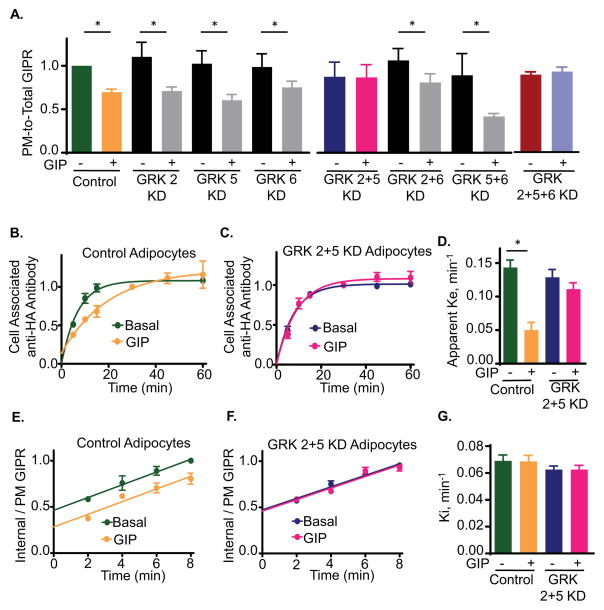
Fig. 2. GRKs 2 or 5 are required for GIP-stimulated GIPR sequestration and slowed recycling.
(A) Quantitation of GIPR PM-to-Total distribution in basal and GIP-stimulated WT (no siRNA), and GRK KD adipocytes. Data are averages of 5 independent experiments ± SD., p≤0.05.
(B) GIPR Exocytosis experiment in WT adipocytes. Data are averages ± SD of 5 independent experiments.
(C) GIPR exocytosis in GRK2+5 double KD adipocytes. Data are average ± SD of 5 independent experiments. Each GRK 2+5KD experiment was accompanied by an experiment in WT adipocytes (shown in panel B).
(D) Exocytic rate constants (Ke) for GIPR in WT or GRK2+5 KD cells calculated from (B) and (C). The data were fit to a single phase exponential rise equation. Data are averages of 5 independent experiments ± SD., p<0.05.
(E) GIPR internalization in WT adipocytes. The internalization rate constants are plotted in panel G. Data are averages ± SD of 7 independent experiments.
(F) GIPR internalization in GRK2+5 double KD adipocytes. Data are averages ± SD of 7 independent experiments. Each GRK2+5 double KD was accompanied by an experiment in WT adipocytes (shown in panel E).
(G) Internalization rate constants (Ki) for GIPR internalization in WT or βA2 GRK2+5 double KD. The Ki were calculated as slopes of straight lines from (E) and (F).
See also Fig S1 and S2.

