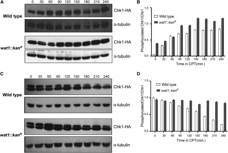
Figure 4.
Chk1 dephosphorylation was delayed in wat1 deletion mutants after release from CPT. (A) Wild-type and wat1-deleted cells containing HA-tagged Chk1 were grown to midlog phase in the presence of 40 µM CPT. Protein lysate from each of the indicated time points was analyzed by western blot using anti-HA antibody; α-tubulin antibody was used as loading control. (B) The graph shows average Chk1 phosphorylation relative to Chk1 protein determined from GelQuant Express Image analysis of data obtained in three independent experiments. (C) Wild-type or wat1-deleted cells containing an HA-tagged Chk1 were grown in the presence of 40 µM CPT for 3 hr, cells were washed and resuspended in fresh medium, and allowed to grow for the indicated time. Protein samples from the indicated time points were analyzed by western blot as described above. (D) Quantitation of Chk1 phosphorylation was plotted as described above. CPT, camptothecin.

