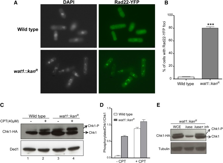
Figure 5.
Deletion of wat1 gene leads to DNA damage. (A) Strains were grown in YEA liquid medium at 25° until midlog phase. The cells were processed for indirect immunofluorescence microscopy using anti-GFP antibody as described in Materials and Methods. (B) Approximately 200 cells for each sample in three independent experiments were counted and the average percentage of cells containing Rad22-YFP foci with SD was plotted. (C) Wild-type and wat1-deleted cells containing HA-tagged Chk1 were grown to midlog phase in the presence or absence of 40 µM CPT. Protein lysate was prepared and probed with anti-HA to visualize Chk1. Anti-Ded1 antibody was used as a loading control. (D) The graph shows average Chk1 phosphorylation relative to Chk1 protein determined from GelQuant Express Image analysis of data obtained in three independent experiments. (E) Protein lysate of wat1-deleted cells was treated with alkaline phosphatase or alkaline phosphatase with inhibitor for 30 min, and probed with anti-HA antibody to visualize Chk1. Anti α-tubulin antibody was used as a loading control. YEA, YE agar; YFP, Yellow fluorescent protein.

