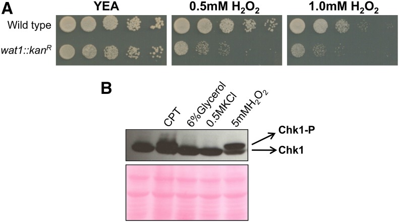
Figure 7.
Sensitivity of wat1 deletion in response to oxidative stress. (A) Ten-fold serial dilutions of logarithmic growing cells were spotted on YE-rich medium, containing the indicated concentrations of H2O2. Plates were incubated at 25° for 3 days. Spotting assay was performed in three independent experiments with similar results. (B) Wild-type cells containing a HA-tagged Chk1 were grown to log phase and treated with 40 µM CPT, 6% glycerol, 0.5 M KCl, or 5 mM H2O2. Protein lysate was analyzed by western blot using anti-HA antibody; ponceau stained gel is shown as a loading control. Experiment was performed three times with similar results. CPT, camptothecin; H2O2, hydrogen peroxide; YEA, YE agar.

