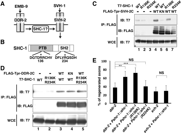
Fig 5. Interactions of SHC-1 with SVH-2 and DDR-2.
(A) The relationship among EMB-9–DDR-2, SHC-1 and SVH-1–SVH-2 in the JNK signaling pathway. (B) Structure of SHC-1. Dark and hatched boxes represent the PTB and SH2 domains, respectively. Essential Arg residues required for binding to phospho-tyrosine in PTB and SH2 domains are indicated by asterisks. (C,D) Interactions of SHC-1 with SVH-2 and DDR-2. COS-7 cells were transfected with plasmids encoding FLAG-Tpr-SVH-2C (WT), FLAG-Tpr-SVH-2C(K767R) (KN), FLAG-Tpr-DDR-2C (WT), FLAG-Tpr-DDR-2C (K554E) (KN), T7-SHC-1 (WT), T7-SHC-1 (R136K), T7-SHC-1 (R234K) and T7-SHC-1 (R136K; R234K), as indicated. Whole-cell extracts and immunoprecipitated complexes obtained with anti-FLAG antibody (IP: FLAG) were analyzed by immunoblotting. (E) Percentages of axons that initiated regeneration 24 hr after laser surgery. Error bars indicate 95% CI. **P<0.01, ***P<0.001. NS, not significant.

