Figure 4.
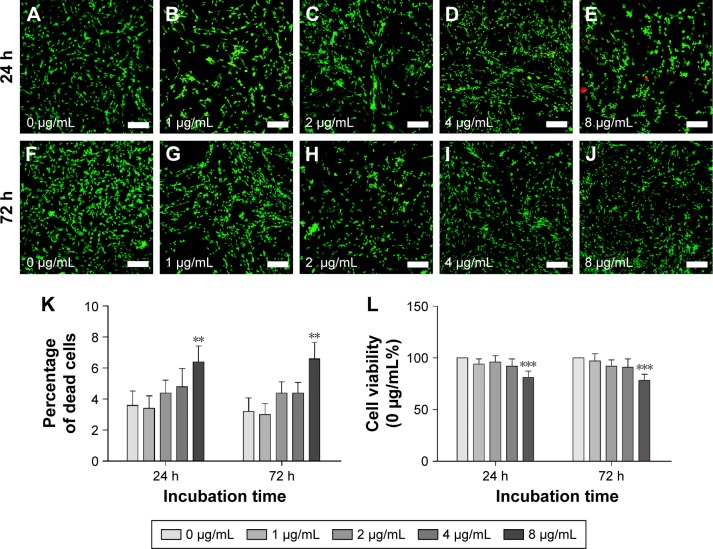
SC cytotoxicity tests of PEI-SPIONs.
Notes: SC cytotoxicity tests of PEI-SPIONs were carried out using live–dead staining and CCK-8 assay. Images of live–dead assay 24 h (A–E) and 72 h (F–J) after SCs were treated with different concentrations of PEI-SPIONs. (K) The percentage of dead cells was examined by live–dead assay. (L) Cell viability was evaluated by CCK-8 assay. Live cells were stained in green, while dead cells were stained in red. Scale bar: (A–J) 100 μm. Graph bars: mean ± SD; **P<0.01, ***P<0.005.
Abbreviations: CCK-8, Cell Counting Kit 8; PEI-SPIONs, polyethylenimine-coated superparamagnetic iron oxide nanoparticles; SCs, Schwann cells; SD, standard deviation.

