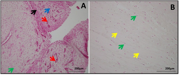
Fig 2. NFR staining in shoulder tendon showing the morphology and orientation.
(A) Group 1 patient (representative of 4 individual subjects in Group 1) with tendinopathy as well as glenohumeral arthritis and (B) Group 2 patient (representative of 4 individual subjects in Group 2) with tendinopathy but without glenohumeral arthritis. The green arrows show tendon cells, red arrows point blood vessels indicating angiogenesis, blue arrows indicate ECM disorganization, black arrows show inflammation and yellow arrow mark normal ECM. The figures were taken in 400x magnification.

