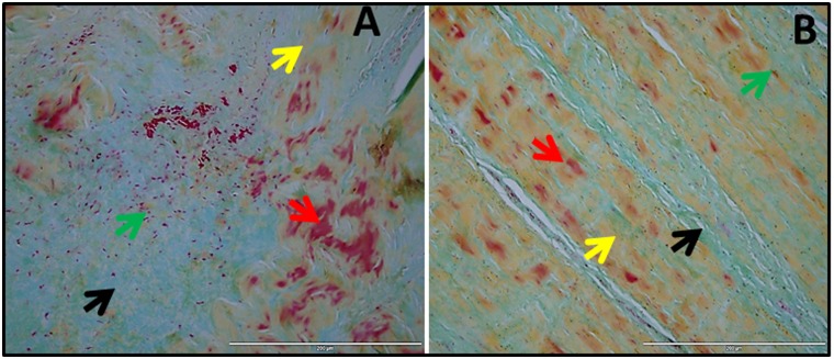
Fig 3. Movat pentachrome staining of shoulder biceps tendons.
(A) Group 1 patient (representative of 4 individual subjects in Group 1) with tendinopathy as well as glenohumeral arthritis and (B) Group 2 patient (representative of 4 individual subjects in Group 2) with tendinopathy but without glenohumeral arthritis. The red arrows indicate fibrosis and muscle fibers, yellow arrows show collagen fibers, green arrows mark tenocytes and black arrows show mucin deposits.

