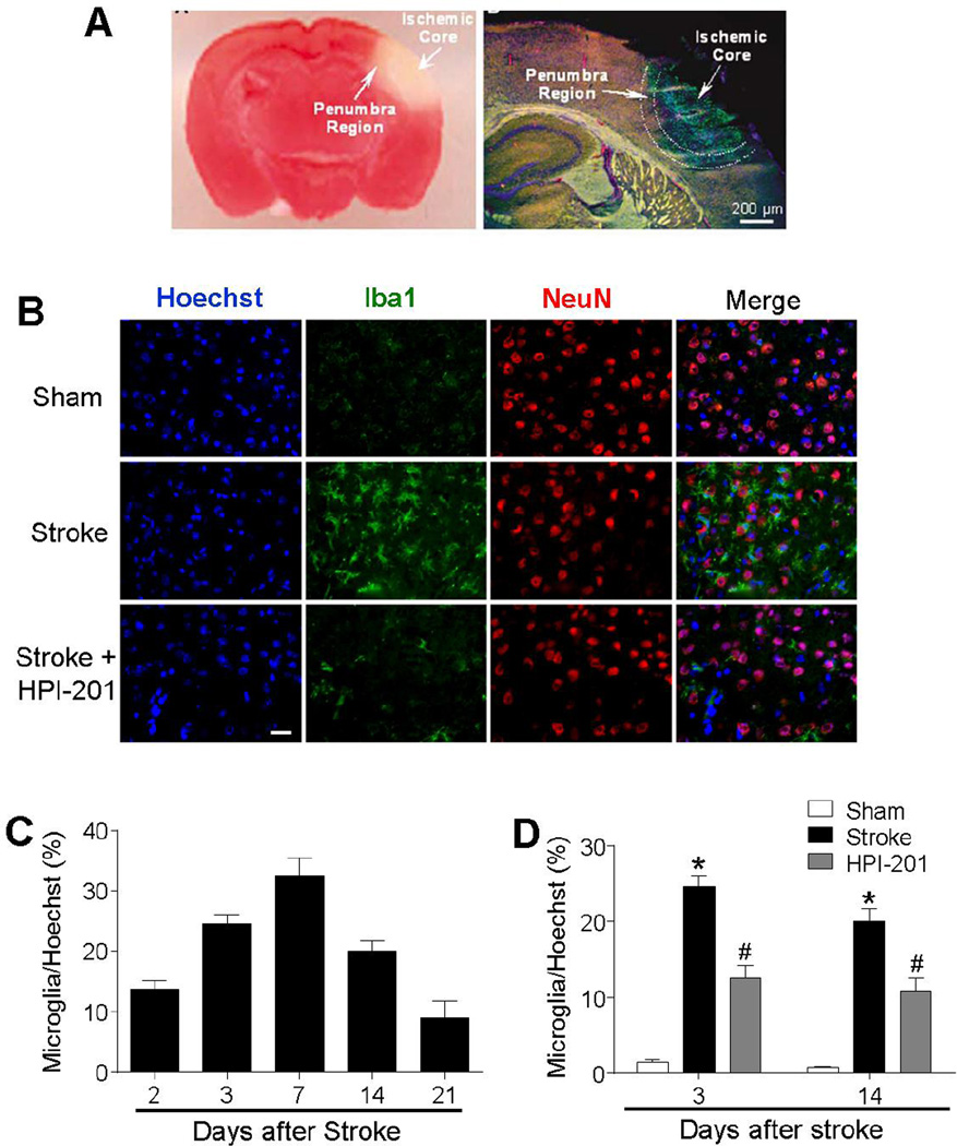
Figure 3. TH reduced microglial activation in the ischemic brain.
Cell type identification and regulation in the peri-infarct (penumbra) region of the post-stroke brain. A. Left: TTC staining 24 hrs after ischemia showing the ischemic core and the peri-infarct region. The pink area between the normal cortex and ischemic core represents the bordering penumbra area. Right: A brain section at lower magnification shows the massive cell death (TUNEL-positive cells of green color) in the ischemic core and scattered cell death in penumbra 3 days after ischemia. Red: Glut-1 staining of vascular endothelial cells, Blue: NeuN staining of neurons. B. Representative images showing immunostaining of Iba1 (green), NeuN (red), and Hoechst 33342 (blue) at 3 days after stroke. C. The time course of microglia recruitment to the penumbra region 2 to 21 days after stroke. D. Effects of HPI-201 and vehicle treatment on microglia recruitment. HPI-201 attenuated the number of Iba1-positive microglial cells at 3 and 7 days after stroke. * P<0.05 versus sham group; #P<0.05 versus stroke group; n=7–8 per group. Scale bars = 20 µm.

